Image Processing for Microscopic Images
![]()

![]()
An Automated System for Parasitic Worm Eggs Detection and Classification on a Motorised Stage Microscope
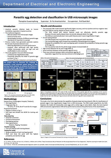
Intestinal parasitic infections remain among the leading causes of morbidity worldwide, especially in tropical and sub-tropical areas with more temperate climates. In this context, we aim to develop a novel cost-effective diagnosis system that automates detection and identification of intestinal parasite eggs from faecal smear samples, that can be used by non-experts. This is a highly interdisciplinary project necessitating this collaborative research proposal.
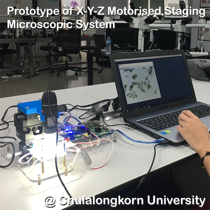
The hardware part, comprising programmable X-Y-Z motorised staging microscopic system, will be developed by researchers from Chulalongkorn University, Mechanical Engineering and Computer Engineering Departments, and the Solimac Automation Co.,Ltd., which is the leading machine vision and automation technology distributor and integrator in Thailand. The algorithmic component, which will automate detection and classification of the parasitic worm eggs from the microscopic images, will be collaboratively performed by researchers from CU's Clinical Microscopy and Computer Engineering Departments, and researchers from Bristol Vision Institute, and Faculty of Life Sciences, University of Bristol who are the experts in medical imaging and machine learning. The system will be tested and validated by personnel at the Phon Phisai Hospital, a district hospital in northeast Thailand.
Posters
Publications
- T Suwannaphong, S Chavana, S Tongsom, D Palasuwan, T H Chalidabhongse, and N Anantrasirichai, Parasitic Egg Detection and Classification in Low-cost Microscopic Images using Transfer Learning, SN Computer Science. 2023
[ Dataset] - N Anantrasirichai, TH Chalidabhongse, D Palasuwan, K Naruenatthanaset, T Kobchaisawat, N Nunthanasup, K Boonpeng, X Ma, and A Achim, ICIP 2022 Challenge on Parasitic Egg Detection and Classification in Microscopic Images: Dataset, Methods and Results, IEEE International Conference on Image Processing. 2022.
[ Dataset] [ Website] - P Mayo, N Anantrasirichai, T H Chalidabhongse, D Palasuwan, A Achim, Parasitic egg detection and classification in USB microscopic images, preprint arXiv:2203.02940, 2022
- T. Suwannaphong, Parasitic egg detection and classification in USB microscopic images, MSc in Biomedical Engineering, 2020
Datasets
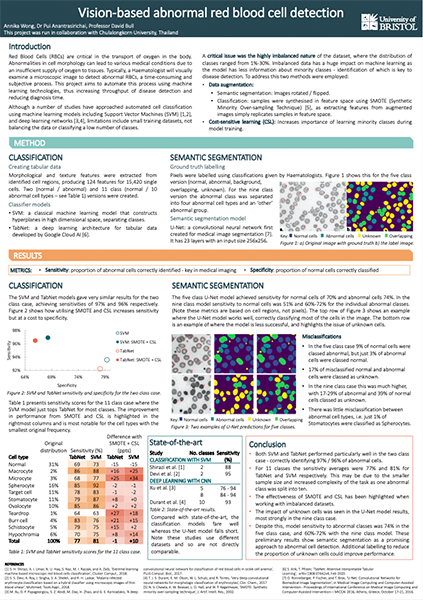
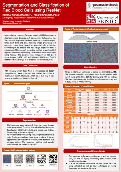
Vision-based Abnormal Red Blood Cells (RBC) Detection and Classification
Identification of abnormalities in red blood cells (RBC) is key to diagnosing a range of medical conditions from anaemia to liver disease. Currently this is done manually, a time-consuming and subjective process. This project aims to automate this process utilising the advantages of machine learning to increase capacity and standardisation of cell abnormality detection and cell type classification. To classify multiple classes with deep learning, imbalance problems are common in medical imaging because normal samples are always higher than rare disease samples. This project presents new methods to segment and classify red blood cells from blood smear images, specifically to tackle cell overlapping and data imbalance problems.
Posters
Publications
- A. Wong et al. Analysis of Vision-based Abnormal Red Blood Cell Classification, arXiv:2106.00389, 2001
- K. Naruenatthanaset et al. Red Blood Cell Segmentation with Overlapping Cell Separation and Classification on Imbalanced Dataset, arXiv:2012.01321, 2000
- A. Wang, Vision-based Abnormal Red Blood Cell Ddetection, MSc in Biomedical Engineering, 2020